


A new screening platform could dramatically speed up the development of precision cancer nanomedicine by identifying nanoparticles capable of delivering therapies directly to mitochondria inside cancer cells.
The method uses DNA “barcodes” to track and compare dozens of nanoparticle designs simultaneously in living tumour models, allowing researchers to rapidly pinpoint the most effective candidates for targeted drug delivery.
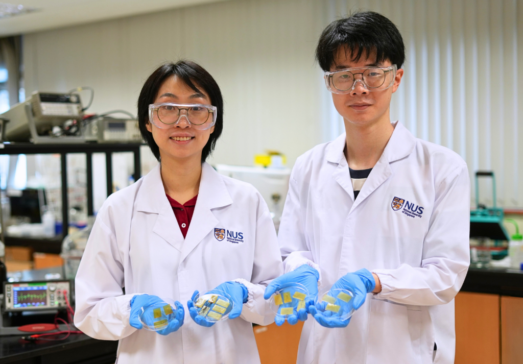
Research led by Assistant Professor Andy Tay (Department of Biomedical Engineering and the Institute for Health Innovation & Technology at NUS) developed the high-throughput platform to systematically evaluate how nanoparticle design, including shape, size and surface chemistry, influences the ability of nanoparticles to accumulate in tumours and reach mitochondria, the energy centres inside cells.
Among the candidates tested, two formulations emerged as standout performers. One, a folic acid-modified cubic gold nanoparticle, achieved 99 per cent tumour regression in preclinical studies when used in a combined treatment involving mitochondria-targeted RNA therapy and mild photothermal therapy.
The study demonstrates how large libraries of nanomaterials can be screened efficiently inside living systems, providing a rational framework for designing nanoparticles that deliver drugs with far greater precision.
The research was published in the journal Advanced Materials.

A barcode system for navigating the body
Mitochondria are attractive targets in cancer therapy because they regulate key processes such as energy production and programmed cell death. Delivering drugs directly to these organelles can disrupt tumour metabolism and trigger cancer cell death. However, nanoparticles must overcome a series of biological barriers before reaching mitochondria: travelling through the bloodstream, entering tumours, penetrating cells and escaping cellular compartments that would otherwise degrade therapeutic cargo.
“Getting nanoparticles to the right place inside the body involves putting them through a complicated obstacle course,” said Asst Prof Tay. “Harnessing DNA barcodes enables us to track many nanoparticle designs simultaneously in living systems and quickly identify which ones can jump through various biological hoops successfully.”
In the study, each gold nanoparticle formulation was tagged with a unique DNA sequence, allowing the researchers to trace its distribution using next-generation sequencing. The team tested a library of 30 nanoparticle designs that varied in shape, size and targeting ligands. After administering the pooled nanoparticles to tumour-bearing preclinical models, the researchers analysed where each design accumulated, from whole organs to specific tumour cell types and ultimately to mitochondria.
This multiplexed approach generated more than 1,000 in vivo data points while requiring around 30-fold fewer preclinical models than conventional one-by-one screening experiments.
The work builds on the team’s earlier study published in November 2024, which first demonstrated the use of DNA barcoding to track nanoparticle biodistribution in tumours. While the previous study compared six nanoparticle designs at the tissue level, this new work greatly expands the library and extends the platform to analyse behaviour at cellular and subcellular scales.
“The results revealed an important insight: nanoparticles that accumulated efficiently in tumours were also far more likely to reach mitochondria,” added Asst Prof Tay. “In other words, successful tumour targeting appears to be a prerequisite for effective subcellular delivery.”
Among the nanoparticle formulations tested, two caught the team’s attention. Large spherical particles modified with folic acid accumulated strongly in tumours, partly due to a protective protein layer that prolonged circulation in the bloodstream. Meanwhile, large cubic nanoparticles entered tumour cells more efficiently through clathrin-mediated endocytosis, a cellular uptake pathway, enabling effective mitochondrial delivery.

A step towards precision nanomedicine
To explore the therapeutic potential of these findings, the researchers tested the cubic nanoparticle formulation in a combined treatment strategy. The particles were engineered to deliver small interfering RNA (siRNA), a molecule that can switch off specific genes, to disrupt mitochondrial gene expression, while also generating heat under near-infrared light through photothermal therapy.
This dual approach produced strong anticancer effects in preclinical studies. When applied together, the treatments led to almost complete tumour elimination after a single dose.
Beyond killing cancer cells directly, the nanoparticles also interacted with tumour-associated macrophages, immune cells that normally support tumour growth. The therapy appeared to shift these cells toward a tumour-fighting state, suggesting the approach may help reshape the tumour immune environment.
“Our findings show that nanoparticle design is not governed by a single factor such as shape or size,” added Asst Prof Tay. “Instead, multiple properties interact in complex ways. High-throughput screening platforms like ours allow us to uncover these relationships and move beyond trial-and-error in nanomedicine design.”
The platform could accelerate the development of precision nanomedicine by enabling researchers to rapidly identify nanoparticle designs suited for specific biological targets. Potential applications include targeted delivery of RNA therapies, gene-silencing treatments and photothermal agents for cancer and other diseases.
Looking ahead, the research team plans to expand the nanoparticle library further and integrate automation and artificial intelligence tools to analyse the large datasets generated by the screening platform. The researchers also aim to extend the method to target other cellular organelles, opening new possibilities for highly specific drug delivery within cells.