


As ovaries age, the tissue surrounding developing eggs becomes stiffer, changing the environment in which they mature. How this physical change affects egg development and quality has remained poorly understood.
Researchers at the College of Design and Engineering(opens in new tab) at the National University of Singapore (NUS CDE) and Department of Biological Sciences at the NUS Faculty of Science(opens in new tab), have now shown how a stiffer environment disrupts the fine cellular connections between eggs and the cells that nourish them. In laboratory experiments using tissue from preclinical models that were cultured in stiff materials, they targeted a signalling pathway in these cells and partly restored egg maturation, even while the surrounding environment remained stiff.
The study was led by Assistant Professor Jennifer Young(opens in new tab) from the Department of Biomedical Engineering(opens in new tab) at NUS CDE and Professor Rong Li(opens in new tab) from the Department of Biological Sciences at NUS. The work was carried out together with the Mechanobiology Institute (MBI), a Research Centre of Excellence hosted at NUS, where Prof Li is its director and Asst Prof Young is a Principal Investigator. The findings were published in Aging Cell(opens in new tab) on 13 August 2026.
“Our findings suggest that some of the effects of ovarian tissue stiffening on egg development may be targetable,” said Asst Prof Young. “This gives researchers promise for developing treatments to preserve egg quality and reduce fertility loss linked to ageing or the build-up of stiff tissue in the ovary.”
A stiffer setting affects developing eggs
An immature egg develops inside a follicle, surrounded by layers of support cells that nourish it. Between the follicles is supporting tissue made up partly of a protein-rich network called the extracellular matrix. This network helps give the tissue its structure and stiffness.
Comparing ovaries from young and aged preclinical models, the team found that the stroma became about 2.5 times as stiff with age. This increase was associated with a build-up of collagen, a structural protein in the matrix. Meanwhile, the follicles themselves showed no age-related change in stiffness.
To determine whether increased stiffness alone could impact egg development, the researchers grew follicles from young preclinical models for seven days in hydrated materials, or hydrogels, designed to mimic the stiffness of young and aged ovarian tissue. In stiffer gels, follicles grew more slowly and support cells multiplied less actively, while the proportion of eggs reaching maturity was about a third of that in softer gels. On top of that, eggs that reached maturity were also more likely to show abnormalities in the meiotic spindle – the structure that separates chromosomes during cell division. These findings showed that a stiff environment could reproduce some features of ovarian ageing in young follicles.
Connection lost
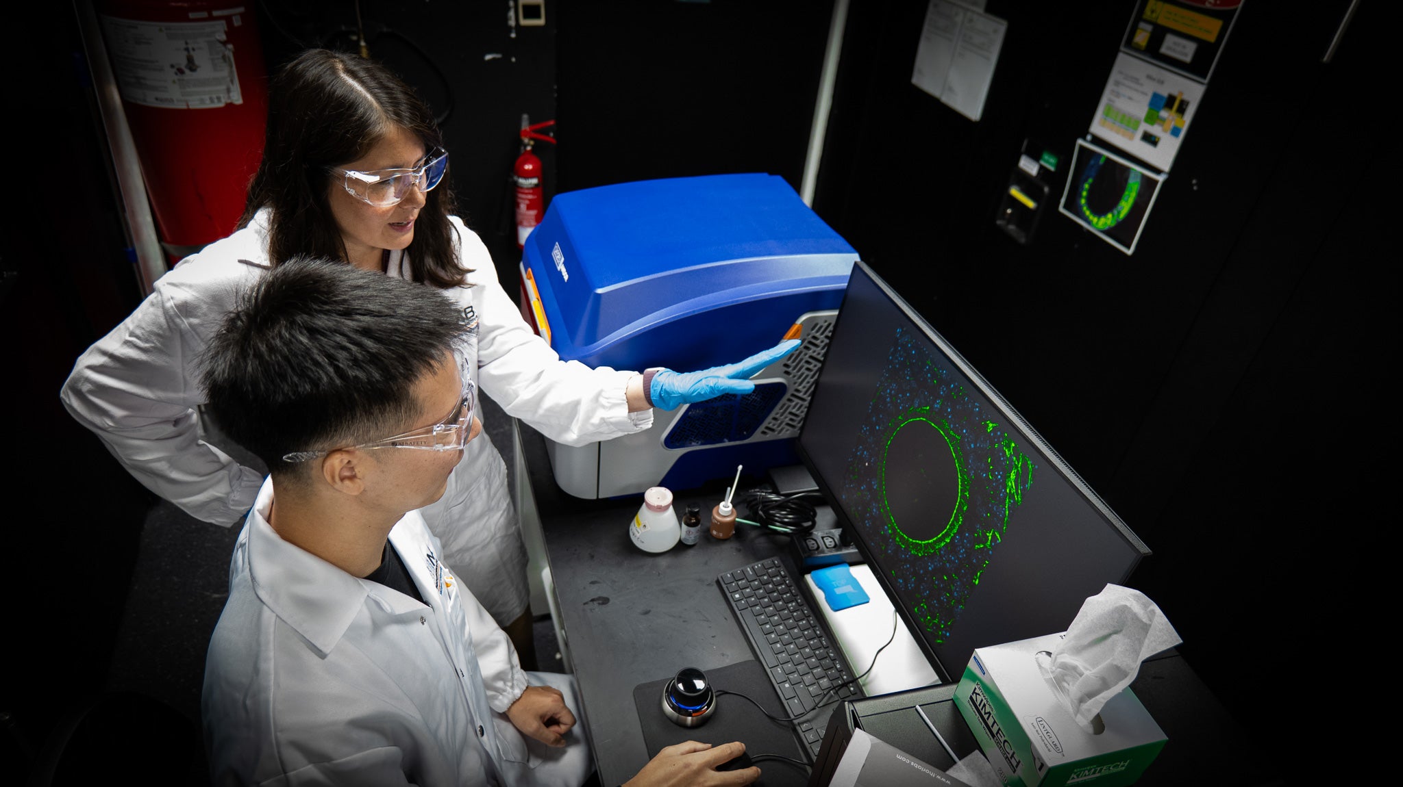
Within each follicle, support cells called granulosa cells extend slender projections, known scientifically as transzonal projections, through the protective layer surrounding the egg. These structures allow the support cells and egg to exchange small molecules and signals needed for development.
Follicles grown in stiffer gels had about 28 percent fewer of these projections per egg than those grown in softer gels. The team also found fewer projections in follicles freshly isolated from aged preclinical models, supporting the link between the laboratory model and changes during natural ageing.
“The egg sits inside the follicle, away from the surrounding tissue,” said and PhD student at the MBI in the laboratories of Prof Li and Young. “Our results help explain how a change in that outer environment can still affect the egg, by disrupting its relationship with the cells that sustain it.”
To understand what was driving this loss, the researchers examined changes in gene activity in the support cells. They traced the effect to the transforming growth factor beta (TGF-β) signalling pathway, which helps these cells form projections that connect them to the egg. In stiff gels, the cells increased activity of the gene, which produces the SMAD7 protein that acts like a molecular brake to dampen TGF-β signalling and impair the formation of these connections.
Improving egg development in a stiff environment
Testing whether releasing this brake could improve follicle function, the team treated cultured follicles with Mongersen, a drug that reduces the production of the SMAD7 protein. They found that suppressing SMAD7 reactivated the TGF-β signalling pathway in the support cells, helping them rebuild connections with the egg even while the surrounding gel remained stiff.
The intervention partially reversed the effects of stiffness on follicle growth and egg development, restoring cellular projections and improving egg maturation. It also improved the function of mitochondria, the energy-producing structures inside eggs, which had been impaired in stiffer gels. Losing these connections could limit the supply of nutrients needed to power the egg’s development.
To advance this work towards potential fertility treatments, the team’s next step is to test whether suppressing SMAD7 can restore cellular connections and improve egg development in follicles taken from older preclinical models. Further studies will also need to establish how follicular cells sense tissue stiffness and respond by increasing SMAD7 production.
In addition, as suppressing SMAD7 only partly restored follicle function, the team posited that other mechanisms may contribute to the effects of tissue stiffening. Therefore, investigating these could reveal additional targets for protecting developing eggs from changes in their surroundings.
“The encouraging finding is that egg development improved even while the surrounding environment remained stiff,” said Prof Li. “This suggests that rather than having to reverse tissue stiffening itself, it may be possible to protect developing eggs by changing how follicle cells respond to that stiff environment.”