

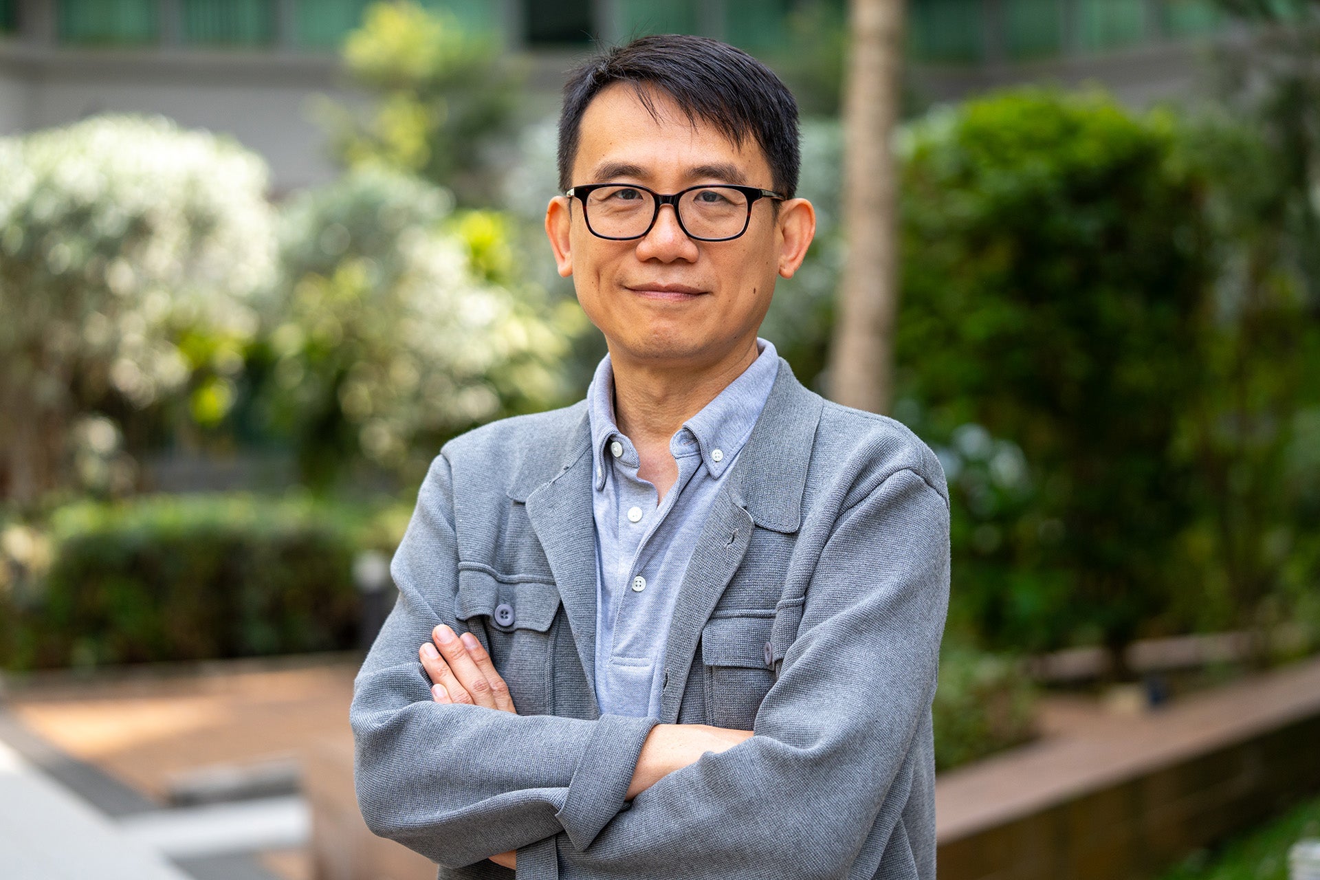
From diagnosing disease to understanding how cells behave, much of biomedical science depends on our ability to see biological structures in ever greater detail. Professor Yang Changhuei (Electrical and Computer Engineering, Biomedical Engineering) has spent his career pushing the limits of what conventional imaging can reveal.
A pioneer in biomedical optics and computational imaging, Prof Yang has developed new ways of combining light, advanced optical systems and computation. His research has driven new approaches to microscopy, produced technologies adopted by industry, and opened new possibilities for imaging and controlling light inside biological tissue.
After more than two decades at the California Institute of Technology (Caltech), where he was the Thomas G. Myers Professor of Electrical Engineering, Bioengineering and Medical Engineering, Prof Yang has returned to Singapore to join NUS, with support from the National Research Foundation’s Returning Singaporean Scientists Scheme (RSSS).
He left Singapore after National Service to study at MIT before building his academic career in the US.
“Throughout my years away from Singapore, I’ve always been grateful for how well it prepared me for my academic career,” he says.
Seeing more with computation
One of Prof Yang’s most influential contributions is Fourier ptychographic microscopy, or FPM, a computational imaging technique first demonstrated by his research group in 2013.
Traditional microscopes face a basic trade-off: looking across a large area generally means sacrificing fine detail, while achieving very high resolution usually limits how much of a sample can be seen at once.
FPM tackles this differently. It captures a series of lower-resolution images under different illumination angles and combines them computationally to reconstruct a highly detailed image across a much wider field of view. It can also digitally correct optical distortions and refocus images.
“Ever since the microscope was invented, making it ‘better’ meant building increasingly complex lens systems to correct blurriness and distortion,” Prof Yang says. “Fourier ptychography was the first method to solve that problem differently: instead of fixing the distortion with more glass and lenses, we fix it with computation.”
The approach became an influential development in computational microscopy, with applications ranging from digital pathology and biological research to high-throughput imaging. Prof Yang’s group has since developed newer methods that can produce high-resolution images without the iterative reconstruction used by FPM.
Looking deeper into biological tissue
Another major strand of Prof Yang’s research tackles how to use light inside biological tissue.
As light travels through tissue it becomes scattered, making it increasingly difficult to focus. Prof Yang and his team pioneered optical time-reversal approaches that exploit the fact that this scattering can, in principle, be reversed.
“There are always new discoveries waiting to be made, ones that can surprise even the most experienced minds in the field.”
Professor Yang Changhuei
By measuring how light has scattered and shaping it so that it effectively retraces its path, researchers can focus light much deeper into tissue. His group later demonstrated the approach by imaging fluorescent structures 2.5 mm deep inside biological tissue.
“When our results first came out, several senior, well-respected scientists told me our results made no sense,” Prof Yang says. “There are always new discoveries waiting to be made, ones that can surprise even the most experienced minds in the field.”
For Prof Yang, such opportunities often emerge where disciplines meet.
“Biophotonics is intrinsically a cross-disciplinary research field that sits between biomedicine and physics,” he says. “It’s rich with opportunities to use good, and often non-obvious, physical insights to develop surprising technologies that let us see more and do more in biomedicine.”
That emphasis on turning scientific insight into useful technology is reflected in the reach of his work beyond the laboratory. Prof Yang holds more than 90 granted patents, with 52 licensed to start-ups and established companies, and was elected a Fellow of the US National Academy of Inventors. Through his new Computational Microscopy, AI and Wavefront Shaping Lab, he aims to build on that record at NUS.
Artificial intelligence is now opening another direction for that work.
Prof Yang’s team recently completed a pilot project using AI to analyse biopsy slides from lung cancer patients and predict which patients would later develop brain metastases, outperforming expert pathologists asked to make the same predictions.
The next step is to understand what the AI is seeing that humans cannot and use that knowledge to rethink the microscope itself. Future systems could be designed specifically to capture information that machines find most useful.
Prof Yang aims to understand how the AI is making these predictions and use that insight to design microscopes that can detect the relevant features more sensitively.
“Instead of designing microscopes to produce images that make sense to the human eye, we’ll be designing them to produce images optimised for AI,” he says. “Ones that may look completely unrecognisable to us, but that machines can read with far greater accuracy.”